Overview
- Brain Morphometry (Computational Neuroanatomy or Neuromorphometry) is the application of computational approaches to quantify anatomical structures in the brain and associated changes, as the brain develops through the lifespan. We can do this using neuroimaging, such as MRI.
- The human brain is a continuously changing and evolving unit, and there are some structural differences among sexes. For example, men have generally higher brain volumes than women. However, this structural difference does not indicate functional superiority.
- A bigger brain may indicate greater cognitive abilities and higher intelligence, but this relationship is not causal and does not make any difference in the daily functioning of typically developing individuals. Brain size/volume is not the only factor that contributes to variability in these abilities among individuals.
- Researchers use measures of brain volume as an indicator of brain health. The brain declines in structure and function as we age, but this is normal! We are interested in changes in brain volume that are not normal, such as those in neurodegenerative conditions, like Alzheimer’s disease.
- While bigger does not necessarily mean better, there are some instances where size does matter, such as in neurodegeneration. And while we cannot jack up our brain to whatever size we want at the gym, we can make small changes in our daily lives that could help us age more gracefully!
Brain Morphometry
In 1919, Walter Dandy introduced pneumoencephalography as an imaging strategy, whereby the cerebral spinal fluid (CSF – fluid surrounding the brain and filling all empty spaces in the head) was replaced with air, which allowed physicians to study the brain directly. As you can imagine, things are done differently these days! To study brain structure in living beings, we primarily use non-invasive neuroimaging techniques, such as magnetic resonance imaging (MRI). Digital data obtained through neuroimaging is then analysed using various mathematical approaches and statistical methods. This allows us to obtain specific brain measurements relating to brain shape, mass, and volume.
A T1-weighted magnetic resonance imaging (MRI) of a normal brain. White matter (light grey), grey matter (dark grey) and CSF (black) can be clearly identified.
Image credit to Dr. Laurent Hermoye. This image is licensed under the Creative Commons Attribution-Share Alike 3.0 Unported license.
Brain Morphometry (also known as Computational Neuroanatomy or Neuromorphometry) is the application of computational approaches to quantify anatomical structures in the brain and associated changes as the brain develops through the lifespan (Grenander & Miller, 1998). Morphometric measurements also provide insight into clinically relevant changes in the brain due to a diseased state or condition which affects brain structure, as well as allow us to see structural changes due to processes like learning and plasticity, language, and even evolution! Some of the commonly used morphometric measures in research are measures of cortical thickness, curvature, area, and brain volume.
The Human Brain
From the moment your brain starts to form in the womb all the way to old age, it constantly changes! Every new thing that you experience or learn influences how your brain will create new networks and pathways. Your brain has as many as 100 billion neurons interconnected by trillions of synapses. In the first year of your life, your brain can form more than a million new connections and triple in size (Kalat, 2018)! For those of us who don’t deal with change very well, it is surprising to hear that our brains change the most throughout our lives, more than any other part of our bodies!
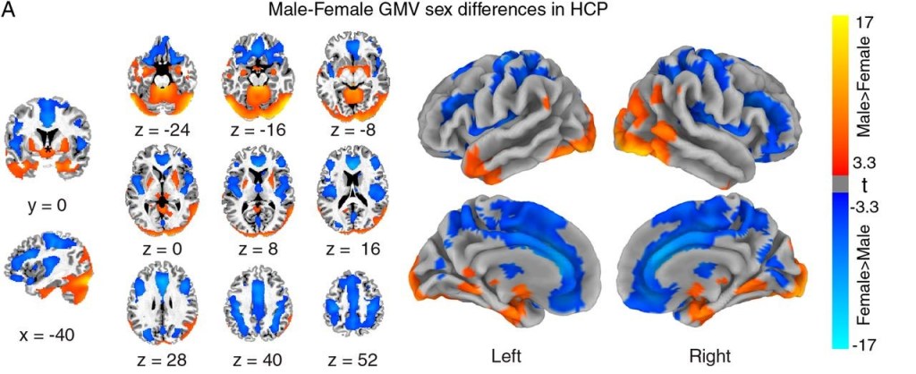
During your lifespan, your brain grows and matures continuously until it reaches its peak size around ages 30-40. We all know too well how irresponsible teenagers can be, but the fact of the matter is, the brain areas responsible for executive functions – the frontal lobes – develop and mature fully much later in life! There also seem to be some differences in brain structure across sexes. Ritchie et al. (2018) recently analysed data from an ongoing large scale biomedical study in the UK, where they examined volumes of 68 brain regions, along with the thickness of the cerebral cortex, and compared them among men and women. They reported that women had higher volumes than men in 10 brain regions, as well as thicker cortices than men, which is associated with better scores in cognitive testing. On the other hand, men had higher total brain volumes, and higher volumes than women in 14 regions, including the hippocampus (implicated in memory and spatial awareness), amygdala (emotions, memory, decision-making), and thalamus (processing and sending sensory information to the rest of the brain).
It is important to note that, like anything related to nature and humans, there are substantial variations in brain volumes and other structural differences among individuals. That is why it is important to remember that structural differences in healthy human beings do not necessarily translate to better or worse functional properties. Therefore, overall brain function is dictated by may distinct factors, and a greater brain volume itself does not necessarily mean better cognitive abilities in a healthy population.
Why Do We Care About Brain Volume?
As tiedmann once said:
“There exists an indisputable connection between the size of the brain and the mental energy displayed by the individual man.”
– Frederick Tiedmann, 1836
A relationship exists between brain size i.e. volume, and cognitive ability among primates – including humans (de Sousa & Proulx, 2014). We have all come across people in our lives, who may require a longer time to understand a new concept, or we ourselves have struggled with learning something new. That’s just how life is! Everyone is different and learns from experience in their own unique way. This is partly because of physiological differences in our brains. For example, while studies on brain volumes and IQ tend to overestimate the relationship between the two concepts, there is some truth to the premise (Pietschnig et al., 2015). Weak to moderate correlations indicate that bigger brained people tend to score higher on IQ tests. While it is very tempting to interpret this in the context of greater cognitive ability, brain volume is not the only factor that may contribute to differences in intelligence (Pietschnig et al., 2015). In fact, a causal relationship has not been established between the two. A big brain could cause high intelligence, or high intelligence could warrant an increase in brain volume, or both could be caused by completely different factors. These associations between structure and cognitive ability are just that – associations!
Brain Volume as a Measure of Brain Health
Perhaps the most important reason why we look at brain volume, is because we view it as an indicator of brain health. Consider aging! As all other systems and parts of our bodies, our brains also decline with age. After the brain reaches its peak in size, it starts shrinking at a rate of 0.5% – 1% per year (Narayanan et al., 2020). This is a normal level of brain shrinkage that occurs with increasing age, affecting both structure and function. Decreases in brain volume translate to more frequent memory lapses and difficulties learning something new, which is pretty much standard the older you get.
However, brain diseases, mental disorders and other conditions affect the brain in an abnormal way, such that changes in volume happen at a higher rate than usual. Brain shrinkage, also referred to as cerebral atrophy is characterized by a loss of neurons and the connections between them, which reduces the size and 3-dimensional volume of the brain. Atrophy can affect the whole brain as one unit, or individual specific brain regions and structures, such as the hippocampus, frontal lobes or visual cortex.
A full lecture program presented for University of California Television (UCTV).
Progressive atrophy is the major event in neurodegenerative diseases, such as dementia and Alzheimer’s disease, Huntington’s disease and Multiple Sclerosis. The rate at which neurodegeneration progresses causes clinically significant changes in brain function. Some examples include substantial loss of memory, an inability to learn, think, plan, organize oneself, problems with speech and understanding language, and an inability to willingly regulate bodily functions.
Problems with the Structure = Function Paradigm
We have been interested in learning more about the intricacies of the human brain since the ancient times, and what makes us different among primates, and unique as individuals. While in general certain structures are related to specific functions in the brain, it does not always mean that individual differences in structure are the cause of functional variation. What do I mean by this?
There has been a notable tendency of scientific racism and neurosexism in research. Structural differences between races have been used to spread the idea that white people are superior to Black people for example, while differences among the sexes have been used to point to male superiority and dominance, and justify harmful stereotypes. The truth is that the quoted structural differences were notoriously over-exaggerated in the past, or even falsified. While we know that there are notable differences among healthy human beings or all ages, races and sexes, these may stem from various environmental and genetic factors.
But the more important point I wish to make is that whatever structural differences do exist, they do not translate to superiority in function. In fact even if we just look at structure alone, it would be very difficult to differentiate between two healthy brains. For example, if a neurologist was looking at two brain scans – one from a healthy man and the other from a healthy woman, it is highly unlikely that they would be able to differentiate between the sexes. Moreover, the individual differences between the man and the woman would not imply that one is smarter or more successful than the other.
The reason we still follow this paradigm of structure = function is to look for answers in pathological conditions. We are still interested in differences between the sexes because statistically speaking males are more frequently diagnosed with some conditions (e.g. autism), and females with others (e.g., major depressive disorder). We want to learn why these trends occur, and we think it might have something to do with the structure, and therefore function of our brains.
We are also immensely interested in slowing or preventing atrophy in neurodegenerative conditions, where the loss of brain tissue directly corresponds to progressively more severe deficits in function. So we do testing and scanning to compare generally heathy brains throughout the life-span with brains affected by these conditions, in order to look for answers. We have also found many associations between certain lifestyle aspects and our brain health as we age. There is an abundant body of research that indicates that a healthy lifestyle consisting of regular exercise, a balanced diet, and lower alcohol consumption among other things, have neuroprotective effects, and aid the aging brain.
In conclusion, while bigger does not necessarily mean better, there are some instances where size does matter, such as when measuring the rate of tissue atrophy in neurodegenerative diseases. And while we cannot jack up our brain to whatever size we want at the gym, we can make small changes in our daily lives that could help us age more gracefully!
Learn More
We will talk about neurodegenerative disorders, including Alzheimer’s in detail in the future, but in the mean time check out the extra resources contained within this page. Hint: look out for linked items!
The World Health Organization places dementia on a high priority list of public health issues, and publishes regular reports and guidelines for risk reduction of cognitive decline, that are a great resource.
Blog Links with Useful Information
- Neuroscientifically Challenged – Neuroscience made simpler: ASSOCIATING BRAIN STRUCTURE WITH FUNCTION AND THE BIAS OF MORE = BETTER
- Neuroscientifically Challenged – Neuroscience made simpler: THE NEUROIMAGING REVOLUTION
- Scientific American: Does Brain Size Matter?
Sources
Cosgrove, K., Mazure, C., & Staley, J. (2007). Evolving Knowledge of Sex Differences in Brain Structure, Function, and Chemistry. Biological Psychiatry, 62(8), 847-855. https://doi.org/10.1016/j.biopsych.2007.03.001
de Sousa, A., & Proulx, M. (2014). What can volumes reveal about human brain evolution? A framework for bridging behavioral, histometric, and volumetric perspectives. Frontiers In Neuroanatomy, 8. https://doi.org/10.3389/fnana.2014.00051
Grenander, U., & Miller, M. (1998). Computational anatomy: an emerging discipline. Quarterly Of Applied Mathematics, 56(4), 617-694. https://doi.org/10.1090/qam/1668732
Kalat, J. (2018). Biological psychology (13th ed.). Cengage Learning.
Liu, S., Seidlitz, J., Blumenthal, J., Clasen, L., & Raznahan, A. (2020). Integrative structural, functional, and transcriptomic analyses of sex-biased brain organization in humans. Proceedings Of The National Academy Of Sciences, 117(31), 18788-18798. https://doi.org/10.1073/pnas.1919091117
Narayanan, S., Nakamura, K., Fonov, V., Maranzano, J., Caramanos, Z., & Giacomini, P. et al. (2020). Brain volume loss in individuals over time: Source of variance and limits of detectability. Neuroimage, 214, 116737. https://doi.org/10.1016/j.neuroimage.2020.116737
Peters, R. (2006). Ageing and the brain. Postgraduate Medical Journal, 82(964), 84-88. https://doi.org/10.1136/pgmj.2005.036665
Pietschnig, J., Penke, L., Wicherts, J., Zeiler, M., & Voracek, M. (2015). Meta-analysis of associations between human brain volume and intelligence differences: How strong are they and what do they mean?. Neuroscience & Biobehavioral Reviews, 57, 411-432. https://doi.org/10.1016/j.neubiorev.2015.09.017
Reite, M., Reite, E., Collins, D., Teale, P., Rojas, D., & Sandberg, E. (2010). Brain size and brain/intracranial volume ratio in major mental illness. BMC Psychiatry, 10(1). https://doi.org/10.1186/1471-244x-10-79
Ritchie, S., Cox, S., Shen, X., Lombardo, M., Reus, L., & Alloza, C. et al. (2018). Sex Differences in the Adult Human Brain: Evidence from 5216 UK Biobank Participants. Cerebral Cortex, 28(8), 2959-2975. https://doi.org/10.1093/cercor/bhy109
Featured Image Credit SpeedKingz/Shutterstock

I used to be able to find good advice from your blog articles.
LikeLike
May I simply say what a relief to discover someone who actually understands what they are talking about over the internet. You actually know how to bring an issue to light and make it important. More and more people ought to look at this and understand this side of the story. I can’t believe you aren’t more popular since you surely possess the gift.
LikeLike
Pretty! This was an extremely wonderful article. Thank you for providing this info.
LikeLike